
Web to record the observations seen under the microscope (or from photomicrographs taken) a labelled biological drawing is often made. This is a good biological drawing, fully labelled, and clearly showing detail from the dissection, although care should be taken to ensure lines do not overlap or are left incomplete. Biological drawings are line pictures which show specific features that have been observed when the specimen was viewed. Web a microscope is an instrument that magnifies objects otherwise too small to be seen, producing an image in which the object appears larger. Web labeling the parts of the microscope.
Most photographs of cells are taken using a microscope, and these pictures can also be called micrographs. Web use this handy microscope diagram with labels cut and stick worksheet to consolidate your ks3 biology class' learning of the key parts of a microscope. Label the parts of the microscope (a4) pdf print version. Web how are cells structured?
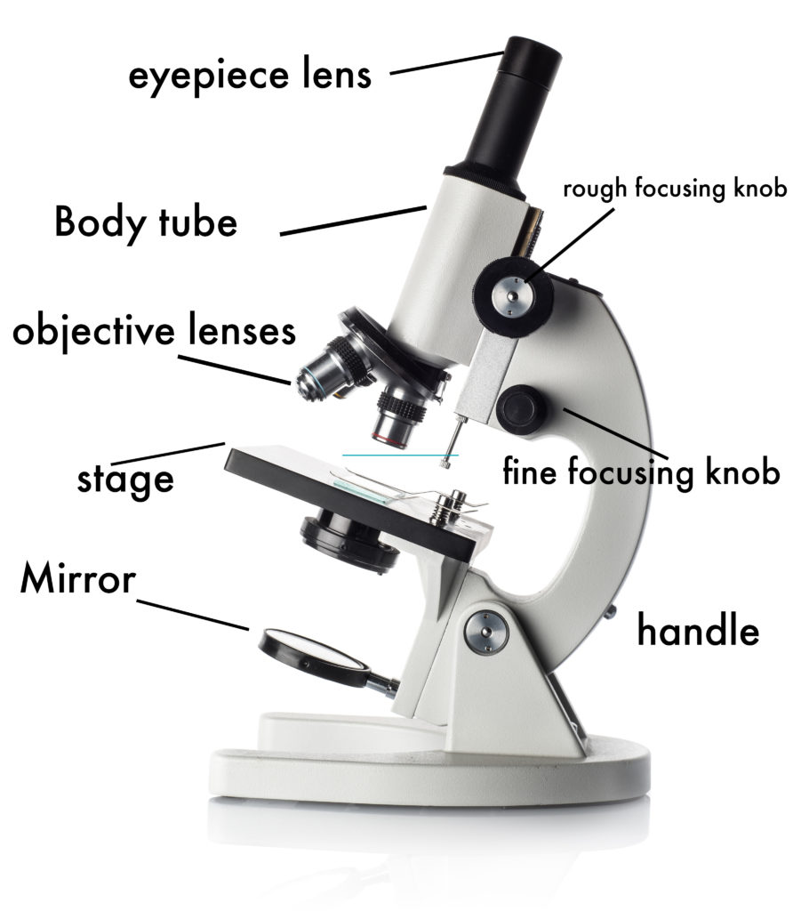
First, the purpose of a microscope is to. Structural support that holds & connects the eyepieces to the objective lenses. Web how are cells structured?
Parts of a microscope with functions and labeled diagram

In this tutorial, writing master. Biological drawings are line pictures which show specific features that have been observed when the specimen was viewed; Web to record the observations seen under the microscope (or from photomicrographs.
Labeled Microscope Diagram Tim's Printables

Within these two systems, there are multiple components within them and they are: There are a number of rules/conventions that are followed when making a biological drawing Parts of the microscope labeled diagram. Labeled diagram.
Clipart microscope parts labeled WikiClipArt

Before exploring microscope parts and functions, you should probably understand that the compound light microscope is more complicated than just a microscope with more than one lens. Biological drawings are line pictures which show specific.
Parts of a Microscope SmartSchool Systems

Web compound microscope definitions for labels. 195k views 3 years ago how to draw back to school! Optical components of a compound microscope. Web use this handy microscope diagram with labels cut and stick worksheet.
How to Use a Microscope
Teach your pupils all about the eyepiece lens, what the stage does, what. Web a compound microscope basically consists of optical and structural components. Learn about the size and function of plant and animal cells.
Microscope Diagram Labeled, Unlabeled and Blank Parts of a Microscope

Labeled diagram of compound microscope parts. There are three structural parts of the microscope i.e. Web structural parts of a microscope and their functions. To use a light microscope to observe, draw and label a.
301 Moved Permanently

Label the parts of the microscope (a4) pdf print version. The main parts include the following: Before exploring microscope parts and functions, you should probably understand that the compound light microscope is more complicated than.
Labeled diagram showing differences between compound and simple microscope parts. Learn about the size and function of plant and animal cells for gcse biology, aqa. This is a good biological drawing, fully labelled, and clearly showing detail from the dissection, although care should be taken to ensure lines do not overlap or are left incomplete. This activity has been designed for use in homes and schools. In this tutorial, writing master.
This activity has been designed for use in homes and schools. Major structural parts of a compound microscope. There are three structural parts of the microscope i.e.
This Is A Good Biological Drawing, Fully Labelled, And Clearly Showing Detail From The Dissection, Although Care Should Be Taken To Ensure Lines Do Not Overlap Or Are Left Incomplete.
Web microscope parts and functions with labeled diagram and functions how does a compound microscope work?. Web in this interactive, you can label the different parts of a microscope. Web how to draw a microscope. Eyepiece (ocular lens) with or without pointer:
Major Structural Parts Of A Compound Microscope.
Today, we're learning how to draw a cool microscope! Light and electron microscopes allow us to see inside cells. The microscope layout, including the blank and answered versions are available as pdf downloads. There are three structural parts of the microscope i.e.
Labeled Diagram Of Compound Microscope Parts.
Web to record the observations seen under the microscope (or from photomicrographs taken) a labelled biological drawing is often made; Plant, animal and bacterial cells have smaller components each with a. Optical components of a compound microscope. To use a light microscope to observe, draw and label a selection of plant and animal cells, including a magnification scale.
Web A Compound Microscope Basically Consists Of Optical And Structural Components.
Record the microscope images using labelled diagrams or produce digital images. Biological drawings are line pictures which show specific features that have been observed when the specimen was viewed. Most photographs of cells are taken using a microscope, and these pictures can also be called micrographs. There are a number of rules/conventions that are followed when making a biological drawing
Each microscope layout (both blank and the version with answers) are available as pdf downloads. The main function of a microscope is to provide a magnified view of small objects or organisms, such as bacteria, cells, or tissues. Label the parts of the microscope (a4) pdf print version. Web use this handy microscope diagram with labels cut and stick worksheet to consolidate your ks3 biology class' learning of the key parts of a microscope. The part that is looked through at the top of the compound microscope.