
The lesson includes educational videos, an interactive quiz, a student checklist, an interactive laboratory powerpoint, and more! Visit the cow’s eye dissection online: Contains detailed instructions, images and an image for labeling the parts of the eye, such as the retina, tapetum, and optic nerve. _____________________ name the three layers that make up the wall of the eyeball. Separate the parts of the eye.
Use your scissors to cut around the. Web this lesson plan describes the cow eye dissection in detail. You should be able to find the sclera, or the whites of the eye. Separate the parts of the eye.
You should be able to find the sclera, or the whites of the eye. This collection details the anatomy of a cow eye. Have students fill out the worksheet about the parts of the eye as you go along.
Cow eye diagrams tipsgamp

(optic nerve, iris, pupil, sclera, cones, rods, cornea, retina, lens and vitreous humor) use a labeled drawing if it is Examine the outside of the eye. Web cow eye dissection 3/6 6. Instructions include an.
Anatomy Of Eye Worksheets Worksheets Cow Eye Dissection

In a preserved cow eye, the lens will most likely have yellowed and become very hard. Web cow eye dissection 3/6 6. You should be able to find the sclera, or the whites of the.
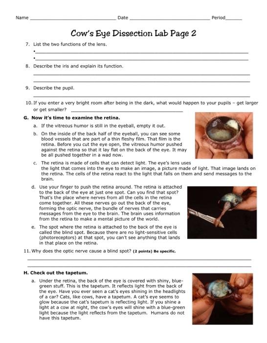
43 cow eye dissection worksheet Worksheet Master
Use your scissors to cut around the. To make the dissection experience for your students Locate the covering over the front of the eye, the cornea. In a preserved cow eye, the lens will most.
Cow Eye Dissection Worksheet Math Sheets For Kids Printable

Follow the directions to dissect a mammalian eye and learn how you see. This tough, outer covering of the eyeball has fat and muscle attached to it. One way to figure out how something works.
Cow Eye Dissection Worksheet Answer Ehydepark

Web learn how to dissect a cow's eye in your classroom. The structures are clear, dissection easy to accomplish and usually kids enjoy the lab. The cornea is made of pretty tough stuff—it helps protect.
Cow Eye Dissection Worksheet Photos

Name three structures that help focus the light rays entering the eye. Examine the outside of the eye. To learn about how your eyes work, you can dissect, or take apart, a cow’s eye. Sds.
Dissection 101 Cow Eye Dissection Lesson Plan PBS LearningMedia

Web this lesson plan describes the cow eye dissection in detail. Web explore learningmedia resources by subject. Web students identify the cornea, sclera, retina, vitreous humor, lens, and optic nerve. You should be able to.
This collection details the anatomy of a cow eye. Middle of the eye, cutting the eye in half. This tough, outer covering of the eyeball has fat and muscle attached to it. Web learn how to dissect a cow's eye in your classroom. If you don’t want to use a scalpel, dissecting scissors will also work!
In a preserved cow eye, the lens will most likely have yellowed and become very hard. Web students identify the cornea, sclera, retina, vitreous humor, lens, and optic nerve. Lab 13 exercise 13.7.1 13.7.
This Tough, Outer Covering Of The Eyeball Has Fat And Muscle Attached To It.
Instructions include an eye diagram, a glossary, and color photos for each step. Web learn how to dissect a cow's eye in your classroom. The lesson includes educational videos, an interactive quiz, a student checklist, an interactive laboratory powerpoint, and more! While the cow was alive, the lens was clear and very flexible.
Lab 13 Exercise 13.7.1 13.7.
Contains detailed instructions, images and an image for labeling the parts of the eye, such as the retina, tapetum, and optic nerve. The worksheet also contains questions and a labeling exercise. Have students fill out the worksheet about the parts of the eye as you go along. Instructions include an eye diagram, a glossary, and color photos for each step.
The Structures Are Clear, Dissection Easy To Accomplish And Usually Kids Enjoy The Lab.
If you don’t want to use a scalpel, dissecting scissors will also work! One way to figure out how something works is to look inside it. Middle of the eye, cutting the eye in half. Web learn how to dissect a cow's eye in your classroom.
Web Learn How To Dissect A Cow's Eye In Your Classroom.
Use your scissors to cut around the. The dissection is very simple and can easily be conducted with younger students. You should be able to find the sclera, or the whites of the eye. The human eye is similar in structure to the eye of other mammals, such as a cow’s.
Locate the covering over the front of the eye, the cornea. Locate the covering over the front of the eye, the cornea. Web learn how to dissect a cow's eye in your classroom. This tough, outer covering of the eyeball has fat and muscle attached to it. This collection details the anatomy of a cow eye.